|
Video capsule endoscopy : definition and technique
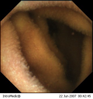
Videocapsule
endoscopy is the latest in cutting-edge technology allowing direct
visualization of the small bowel which could previously only be seen
by surgical techniques. The capsule does provide the unique ability
to film the lining of the small intestine well beyond the reach of
the standard endoscope or enteroscope which at best may only examine
the first 3-6 feet of the small intestine. However, the capsule will
examine the entire length (approx. 20 ft) of the small bowel. Early
studies have shown a 60–70% success rate in identifying the source
of bleeding in the intestinal tract where standard diagnostic tests
had failed.
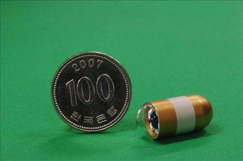
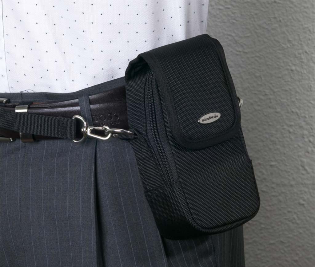
The capsule is a small camera about the size of a vitamin capsule. It emits a light and takes two pictures per second as it traverses the gastrointestinal tract. The pill is easily swallowed after a modified diet on the previous day and an overnight fast. Receiving sensors are taped to the abdomen and the receiver and battery pack are worn around the waist. Once the setup is confirmed to be functional (approximately after 20 minutes), the individual is free to leave the office and participate in most of their usual activities.
The receiver and battery pack are removed in 20
minutes time. The capsule will eventually be excreted in the stool
and is not reusable. It is important to avoid any areas with strong
magnetic fields while the capsule is transmitting, as a magnetic
field will shut it off. One also must refrain from going into an MRI
machine until the capsule passes into the toilet, otherwise serious
injury to the gastrointestinal tract may occur. Finally, the
recorded images are downloaded to a computer where they are reviewed
by a specialized nurse and gastroenterologist. Your treating
physician will be informed within 72 hours and receives an official
report including a medical advice.
|
|||
|
|